
|
|
|||||
|
|
|
|
|
|
|
YOUR LATEST ISSUE ABOUT MRCP PACES IS HERE!
Look at this gentleman fundus and proceed.( Station 5)
( Photo Source: Atlas of Clinical Diagnosis)
Discussion:
This is a popular case in MRCP PACES station 5 ( fundoscopy station). We have discussed about Retinitis Pigmentosa in previous issue ( Issue 22). This is an easy case because you notice there is presence of optic disc swelling with lost of margin. You must always remember that papilloedema is associated with enlargement of blind spot ( this is a popular question examiners ask during your MRCP PACES exam). Another important topic you must remember about papilloedema during your PACES examination is the common causes. Remember that papilloedema is an important diagnostic sign for either systemic illness, intracranial or CSF ( central spinal fluid) problems.As a practising doctor, you notice that you find papillodema in patients with intracranial pressure which can be due to meningitis, intracranial bleeding, or even benign intracranial hypertension. However, always remember about hypertension.
During your MRCP PACES examination, examiners like to ask you other causes of papilloedema which I think you must always remember. There are a few systemic illness thatcan lead to papilloedema. You find that these causes are mailnly metabolic causes such as carbon dioxide retention, thyroid eye disease and lead poisoning.
Other uncommon causes of papilloedema are spinal cord tumour and Guillan-Barre syndrome.
Always try to find out the underlying cause of papilloedema in your patient duting your MRCP PACES, therefore at least suggest to your examiners that you would like to examine your patient's cranial nerves, blood pressure and thyroid status.
Common questions examiners would ask you,
1) What do you know about benign intracranial hypertension?
2) How do you differentiate papillitis from papilloedema?
3) How do you differentiate papilloedema cased by accelerated hypertension from intracranial tumour?
Extra points:
1) Get more information about Papilloedema in Atlas of Clinical Diagnosis by M Afzal Mir
| Case ID:2 | Created: 15 August 2007 |
Interesting Images in Clinical Medicine
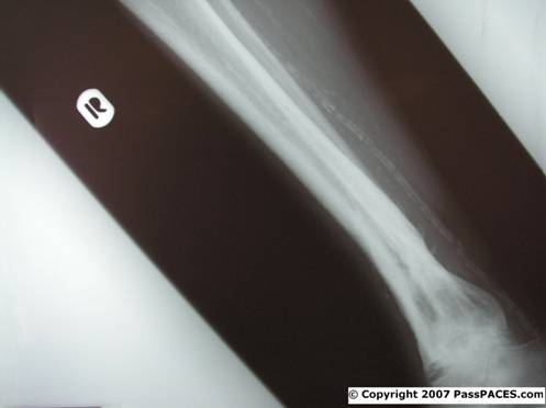
Case Summary:
This is a Xray I took for one End Stage Renal Failure patient undergoing haemodialysis for the past 10 years. He complains of bone pain over hsi shin. What do you notice in this Xray and why? Send your quick comment to us in our blog area!
To see previous issues, click here! To send a quick comment, click here!