
|
|
|||||
|
|
|
|
|
|
|
YOUR LATEST ISSUE ABOUT MRCP PACES IS HERE!
Examine this gentleman's cranial nerves.
Discussion:
As I mentioned in MRCP Issue 11, this is a popular case in your MRCP PACES examination. You can send your comments to our shoutbox area. Since I have discussed before in previous issue how to approach a case with 7th nerve palsy, I think I would talk more about facial nerve in this issue.
First point to remember about facial nerve, it contains three components, namely sensory, motor and parasympathetic. The nerve consists of two roots, nervus intermedius that carries sensory and parasympathetic fibres and another root that composed of motor fibres.
Although you will be dealing with mainly motor components in your MRCP PACES examination, remember that the sensory fibres of the facial nerve supply 1) anterior two-third of the tongue, 2) floor of the mouth and palate, and 3) cutaneous sensation from part of the external ear.
Whereas for the motor fibres, they are distributed to the muscles of facial expression, platysma, stylohyoid, the posterior belly of the digastric muscle and the stapedius muscle of the middle ear. One extra point to remember, those controlling motor neurones which supply the muscles of the upper face ( frontalis, orbicularis oculi) are distributed bilaterally, therefore, uinlateral upper motor neurone lesions, only give rise to paralysis of the lower portion of facial muscles.
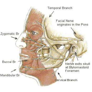
( Picture Source: Bell's Palsy Infosite )
As for the parasympathetic component, it supplies the submandibular, sublingual and lacrimal gland and the nasal and oral mucous membranes.
In this case, it is obvious that you are dealing with Bell's palsy ( first described by Scottish anatomist Charles Bell). You notice the following 1) paralysis of the upper and lower portions of facial muscles, 2) absent corneal reflex and if you examine further, you may find hyperacusis of the affeced side.
Remember that in a neurology station, the examiners are interested in two things ( what is the lesion?- pathological diagnosis and where is the lesion?- anatomical diagnosis), if possible, if you find a lower neurone lesion of 7th nerve, you have to localise the lesion.
Common questions examiners would ask you,
1) How do you localise the site of lesion if you find lower motor lesion of 7th nerve palsy?
2) How do you manage this patient?
3) What is the diagnosis if you find vesicles over the ear of this patient?
Extra points:
1) Get more information about Facial nerve anatomy in Neuroanatomy by A R Crossman
| Case ID:2 | Created: 22 September 2007 |
Interesting Images in Clinical Medicine

Case Summary:
This is a CT scan with contrast of a young patient who presented to our hospital with fever and seizure. What do you notice in this CT brain and what is your diagnosis? Send your quick comment to us in our Forum!
To see previous issues, click here! To send a quick comment, click here!